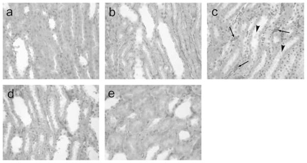
Figure 7.
Histology of renal tubular system in rats toploading with NanoHb (a), LactRing (b), SFHb (c), PolyHb (d) or RBC (e). Under light microscope, in the groups a, b, d and e, the tubular system showed normal, no protein or other sediments within the tubular lumens. However, in the rats infused with SFHb (d), the proximal tubular necrosis was observed (arrows), and necrotic cell debris presented in the lumens of proximal tubules (arrow head), showing the ongoing necrosis in the kidney, although 3 weeks after SFHB injection. H&E staining, ×400.

