Figure 1.

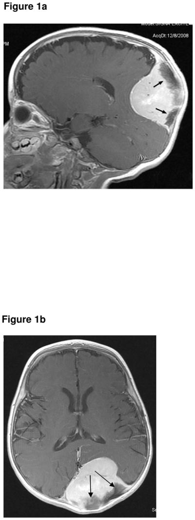
Figure 1a. Sagittal, contrast-enhanced, T1-weighted image (1.5T, TR 650, TE 15) showing a solid, intensely enhancing, large, intracranial, extra-axial, tumour. The dark structures (arrows) are spiculated bone, a known feature of MNTI.
Figure 1b. Axial, contrast-enhanced, T1-weighted MR sequence (1.5T, TR 700, TE 14) showing the large enhancing tumour compressing but not invading the adjacent brain. The dark structures (arrows) are spiculated bone, a known feature of MNTI.
