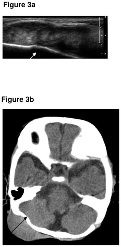
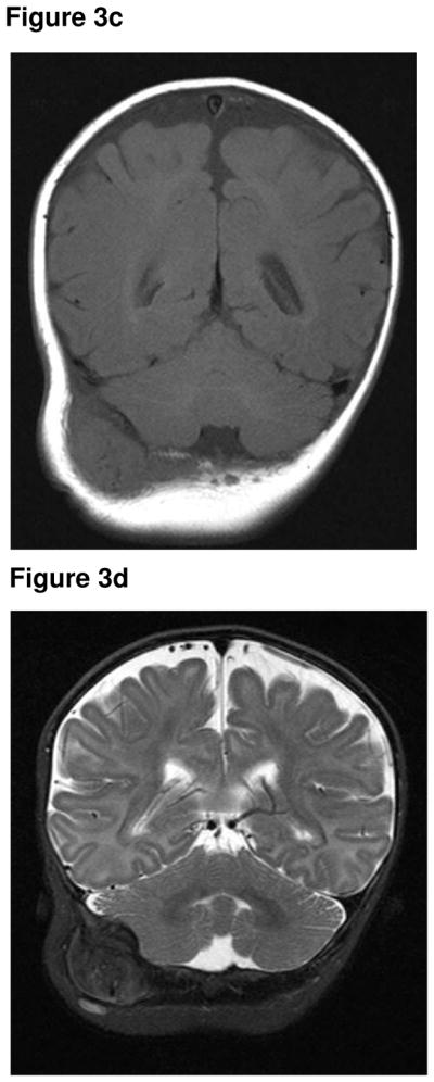
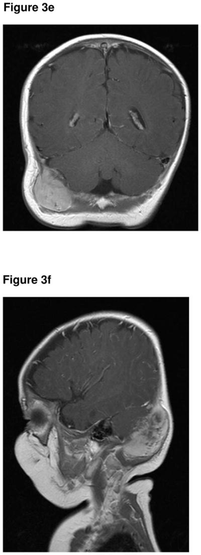
Figure 3.


Figure 3a. Ultrasound demonstrates a well-defined, heterogenous solid lesion adjacent to the right occipital bone (arrow).
Figure 3b. Axial CT of the brain demonstrated a well-defined, isodense lesion closely applied and indenting the right side of the occipital bone (arrow). No bone destruction or intracranial extension.
Figure 3c. Coronal T1-weighted MR sequence (1.5T, TR 561, TE 13) showing an isointense lesion, which has increased in size from the CT.
Figure 3d. Coronal T2-weighted MR sequence (1.5T, TR 6180, TE 115) showing a inhomogenously hypointense lesion.
Figure 3e. Coronal post contrast T1-weighted MR sequence (1.5T, TR 561, TE 13) showing marked contrast enhancement of the lesion.
Figure 3f. Coronal post contrast T1-weighted MR sequence (1.5T, TR 561, TE 13) showing marked contrast enhancement of the lesion. No intracranial extension.
