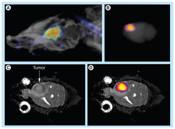
Figure 5. Macroscopic fluorescence imaging.
(A) The in vivo fluorescent image of the rat head overlayed on an x-ray image shows the presence of the agent in the U87 tumor in the brain. (B) The ex vivo fluorescence image of the whole brain also detected the agent in the brain (fluorescent image was overlayed on x-ray image of the whole brain). (C) The coronal MRI shows the location of the U87 tumor. (D) The ex vivo fluorescence image was also overlayed on the MRI to show that the agent was located in the U87 glioma. The anatomy of the brain was correlated between MRI and x-ray images.

