Figure 2.
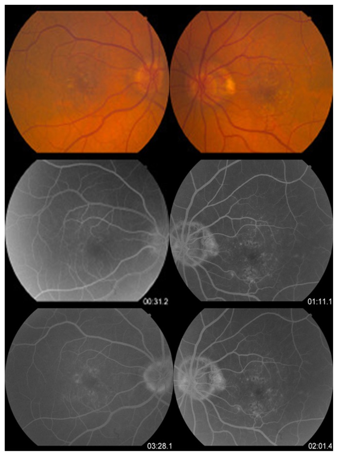
Fundus color photographs and early and late phases of fluorescein angiography.
Notes: In the color photographs, there is evidence of pigmentary changes at the level of the retinal pigmented epithelium and drusen formation in both macula. Fluorescein angiography shows early and late hyperfluorescence probably due to the aforementioned changes.

