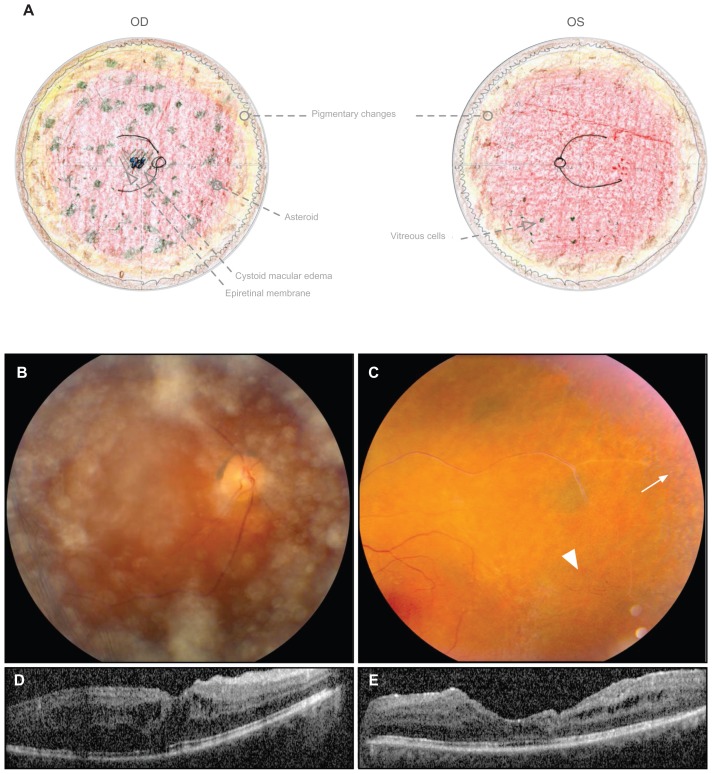
Figure 2.
Twin A displays stage III ADNIV at the age of 49 years. (A) Fundus drawings record 1+ epiretinal membrane, asteroid, and cystoid macular edema OD; attenuated vessels and vascular remodeling OS; and peripheral pigmentary changes and mild vitreous cells OU. (B) Fundus photograph OD shows asteroid in the vitreous. (C) Fundus photograph OS shows a sheathed vessel, pigmentary changes (arrow) in the peripheral macula, and vascular remodeling (arrowhead). (D and E) Optical coherence tomography of the foveas shows cystoid macular edema.
Abbreviations: ADNIV, autosomal dominant neovascular inflammatory vitreoretinopathy; OD, right eye; OS, left eye; OU, both eyes.

