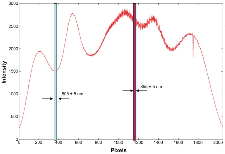
Figure 2.
Spectrum of the light source of the slit-lamp adapted ultrahigh-resolution optical coherence tomography.
Notes: Calibrated pixel locations on the charge-coupled camera for wavelengths of 805 ± 5 nm and 855 ± 5 nm were identified. The fringe from these pixels was processed for calculating the optical intensity of the major retinal artery and vein.

