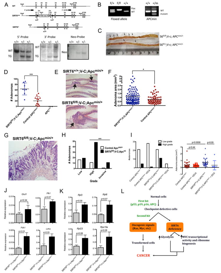
Figure 7. SIRT6 functions as a tumor suppressor in vivo.

A) Strategy to target the SIRT6 locus (upper panel). Southern blot (5’, 3’ and Neo probes) of KpnI-digested genomic DNA showing the targeted allele in the heterozygous cells (+/-) (lower panels). B) PCR showing the presence of the SIRT6 floxed allele (left) and the mutant APC allele (right). C) Representative image of a intestine section from SIRT6fl/fl;V-c;APCmin/+ and SIRT6fl/+;V-c;APCmin/+ mice. Arrows indicate the presence of polyps. D) Adenoma number in the intestines of mice of the indicated genotype. E) H&E staining showing the adenoma size in the indicated mice. F) Adenoma area in SIRT6fl/fl;V-c;APCmin/+ and Control;APCmin/+ mice. G) Representative image and (H) quantification of the grade of the tumors in the indicated mice. I) Grade (righ panel) and area (left panel) of the adenomas in SIRT6fl/fl;V-c;APCmin/+ and Control;APCmin/+ mice untreated or treated with DCA (5g/L). J) and K) Expression of several glycolytic and ribosomal genes in adenomas (n=3) of SIRT6fl/fl;V-c;APCmin/+ and SIRT6fl/+;V-c;APCmin/+ mice (error bars indicate SEM). See also Figure S7.
