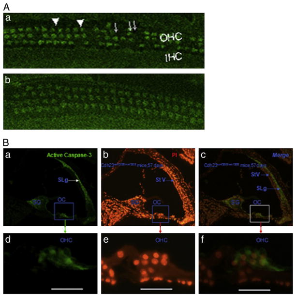
Fig. 7.
Cdh23nmf308/nmf308 mice with AHL exhibited OHC cell apoptosis. (A) Laser confocal inspection of OHCs of Cdh23nmf308/nmf308 (a) and Cdh23ahl/ahl mice (b) at 42 days of age. (B) Detection of apoptosis by caspase-3 and PI staining in fixed sections from Cdh23nmf308/nmf308 mice at 57 days of age. Caspase-3 positive cells were distributed in the organ of Corti (OC), SG and SLg (a), especially in the cytoplasm of OHCs (d). SG and OHC were overlapping with both caspase-3 and PI positive cells. In contrast, the SLg only expressed cas-3, while StV was only stained by PI (c, f). Scale bar: 20 μm.

