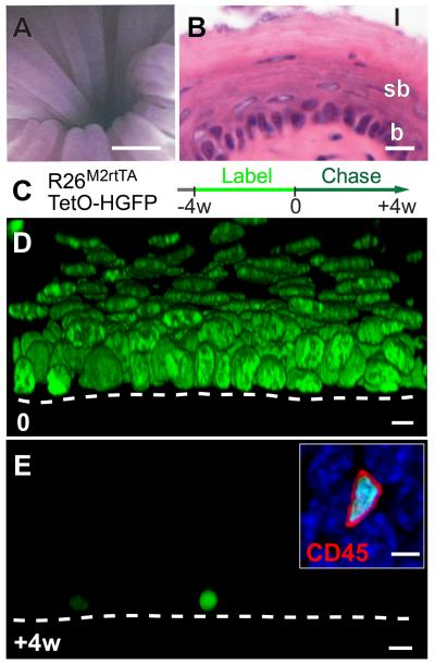
Fig. 1. Esophageal epithelium contains no slow-cycling epithelial cells.
A: Micro-endoscopy showing esophageal lumen, scale bar approx. 500μm.
B: Section of epithelium, basal layer (b), suprabasal layers (sb) and lumen (l), scale bar 10μm.
C: Protocol. Adult Rosa26M2rtTA/TetO-HGFP mice treated with doxycycline (DOX) express HGFP (green). Following DOX withdrawal, HGFP is diluted upon cell division, except in slow-cycling cells.
D, E: Rendered confocal z stacks, showing HGFP (green) at time 0, D, and after 4w chase E (Scale bar 10μm). Dashed line indicates basement membrane. Inset shows CD45 (red) staining in HGFP retaining cell at 4w (DAPI, blue; Scale bar 5μm).

