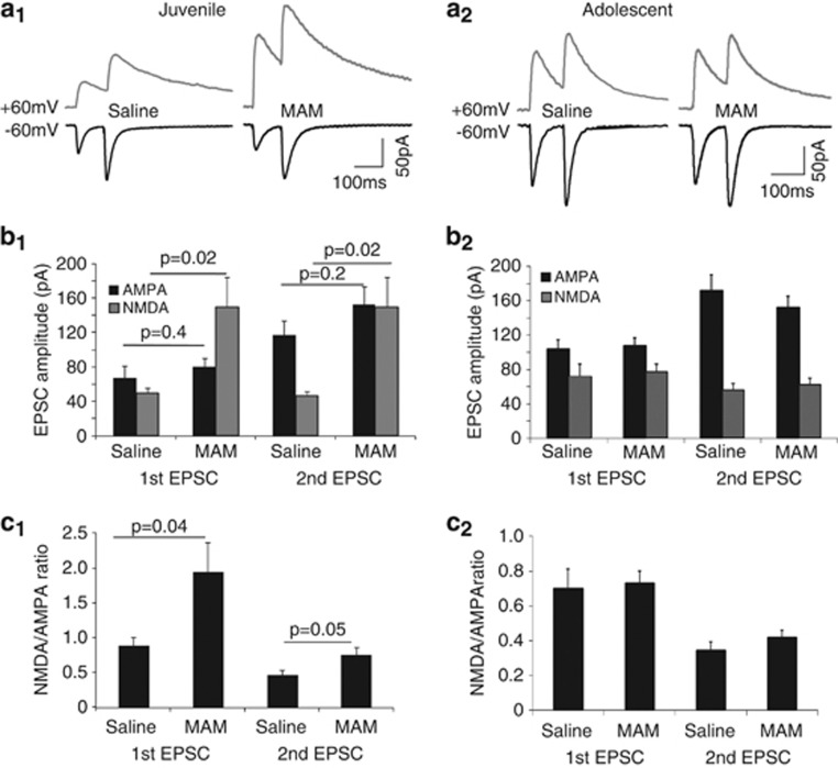
Figure 3.
NMDA–EPSC amplitudes are increased in juvenile MAM-exposed rats, but normalized by adolescence. (a1 and a2) Representative paired-pulse traces of stratum radiatum-evoked EPSCs at −60 and +60 mV from CA1 cells of a juvenile (a1) and (a2) adolescent saline and MAM-exposed animal. (b1 and b2) Summary histograms show that NMDA–EPSC amplitude is significantly increased in cells from juvenile MAM-exposed animals (saline, n=7, MAM n=9, p=0.02 and 0.02, respectively), while AMPA–EPSC amplitude is not changed (saline, n=7, MAM n=9, p=0.40 and 0.42, respectively). In contrast, both NMDA–EPSC and AMPA–EPSC are not different between adolescent saline and MAM animals (saline, n=7, MAM n=8, p=0.7 and 0.6, respectively). (c1 and c2) Summary histograms show a significantly increased first NMDA/AMPA ratio in cells from juvenile (saline, n=7, MAM n=9, p=0.04), but not in neurons from adolescent MAM-exposed animals (saline, n=7, MAM n=8, p=0.8).

