Figure 4.
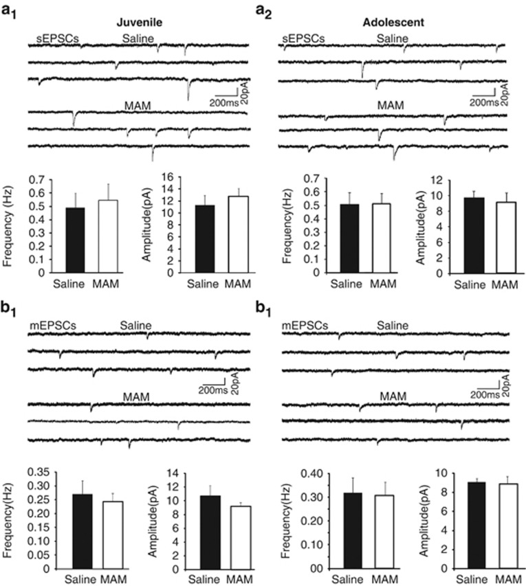
AMPA receptor-mediated currents are not affected by MAM in either juveniles or adolescents. (a1 and a2) Sample traces (upper panels) and summary histograms (lower panels) of sEPSCs recorded at −70 mV in CA1 pyramidal neurons from juvenile (a1) adolescent (a2) saline and MAM-exposed animals. Summary graphs show no change in sEPSC frequency or amplitude with MAM exposure for juvenile (p=0.74, 0.48, respectively, saline n=7, MAM n=8) or adolescent animals (p=0.9, 0.5, respectively, saline, n=7, MAM n=9). (b1 and b2) Sample traces (upper panels) and summary histograms (lower panels) of mEPSCs recorded at −70 mV in the presence of TTX in CA1 pyramidal neurons from juvenile (b1) and adolescent (b2) saline- and MAM-exposed animals. MAM exposure did not alter the frequency or amplitude of mEPSCs for either age group (juveniles, p=0.55, 0.33, respectively, saline n=6, MAM, n=9; adolescent, p=0.7, 0.8, respectively, saline n=6, MAM n=7). These data indicate that there is no change in functional AMPARs at synapses in juvenile or adolescent MAM-exposed animals.

