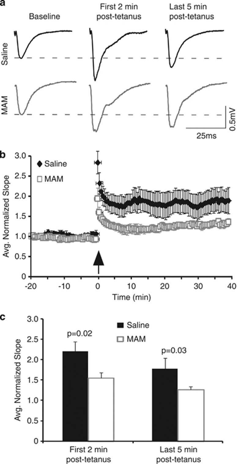
Figure 5.
LTP is impaired in slices from juvenile MAM-exposed animals. (a) Representative fEPSPs recorded in the CA1 region of the hippocampus evoked by stimulation of Schaffer collaterals during baseline recording and following high-frequency stimulation. Recordings were conducted in slices from animals aged P15–21. (b) Summary graph shows the average-normalized slope of fEPSPs in saline and MAM animals during baseline recording and following high-frequency stimulation. (c) Summary histogram shows that both post-tetanic potentiation and LTP are significantly attenuated in MAM-exposed animals (first 2-min post-tetanus, p=0.02; average last 5-min post-tetanus, p=0.028, saline n=6 and MAM n=12).

