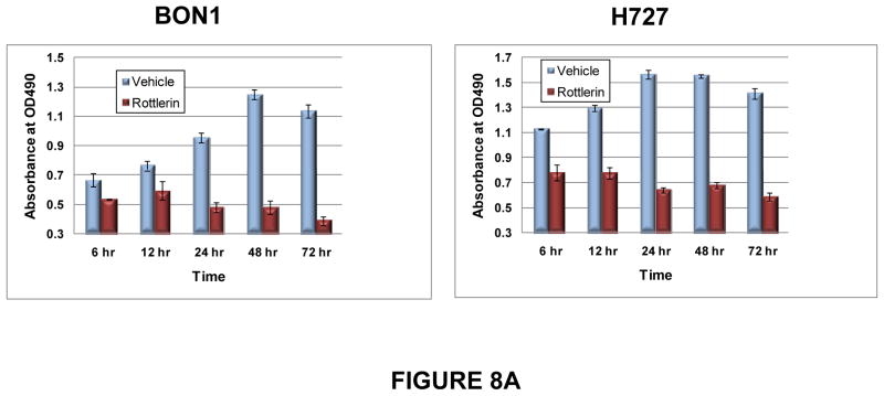
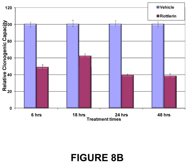
Figure 8. Duration of exposure to PKCδ inhibitors needed to inhibit tumor cell proliferation and clonogenic potential.
(A) BON1 and H727 cells were grown to 30% confluence and then exposed to vehicle or to rottlerin (10 μM) for 6, 12, 24, 48 or 72 hr. Media without inhibitor was replaced and cell numbers were estimated by MTS assay at 24, 48 and 72 hr. Shown here are the results at 72 hr of culture after each washout interval. Error bars represent SEM. Differences in proliferation between rottlerin- and vehicle-treated cultures became statistically significant (p < 0.01) by 24 hr of exposure, and remained significant for all longer periods of exposure. (B) Effects of PKCδ inhibitor on tumor cell clonogenic capacity. H727 cells were grown to 30% confluence and then exposed to vehicle or rottlerin (10 μM) for 6, 12, 24, 48 or 72 hr. Viable cells were enumerated and re-plated in media without inhibitor, and colony numbers were quantitated 96 hr later. Error bars represent SEM. P values for comparison of DMSO control and rottlerin effects on clonogenic capacity reached significance (p=0.0051) at 6 hr of exposure and remained significant for all subsequent exposure times.


