
Figure 2.
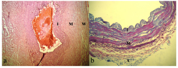
Histological section of HUV and SV. The wall may be divided into Intima (I), Media (M), and Adventitia (A) or Wharton jelly (W). a) Haemotoxylin and Eosin (HE) stained section of HUV. Layers M and W are not uniform (magnification × 100) but thicker than that of SV. b) Van Gison’s stained section of SV. Collagen fibers are blued leafs while red strings stand for smooth muscles. Adventitia is mainly composed of collagen fibers (magnification × 400).