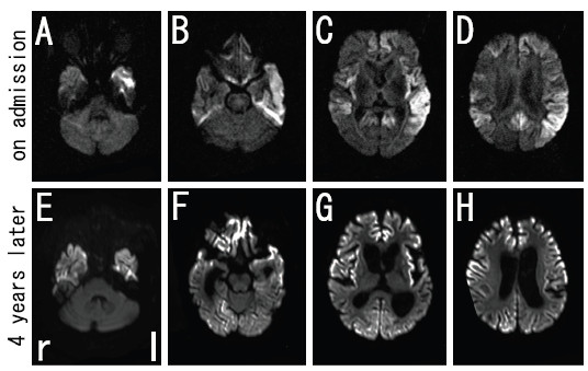
Figure 1.
DWI images of brain MRI in the present case. Increased signal intensity in the bilateral frontal, temporal, and parietal cerebral cortex with left dominancy, except for occipital lobe, was observed on admission with axial imaging (A-D). Note that the high-signal region spread to the occipital cerebral cortex at four years after the onset (E-H).

