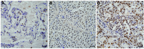
Figure 2.
Immunohistochemical staining of Ku80 in lung adenocarcinoma and adjacent nontumor lung tissues. (A) Ku80 staining was weak in nontumorous lung tissue, (B) low level of expression of Ku80 in lung adenocarcinoma and (C) high level of expression of Ku80 in lung adenocarcinoma. Ku80 staining was brown and predominantly nuclear in the samples examined. Magnification × 400, scale bar 50 μm.

