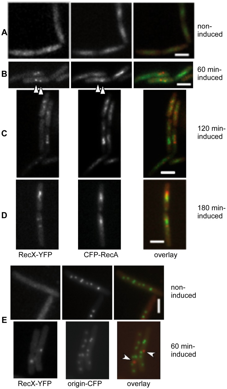
Figure 4. B. subtilis RecX-YFP has a similar subcellular position to CFP-RecA threads and does not co-localize with sites of DSBs.
(A) RecX-YFP, CFP-RecA and overlay in exponentially growing cells. (B–D) Cells at 60 (B), 120 min (C) and 180 min (D) after addition of 0.15 µM MMC. Shown is the corresponding RecX-YFP or CFP-RecA fluorescence, and an overlay of both signals (RecX in red, RecA in green). White triangles indicate examples of colocalization at 60 min. (E) Fluorescent microscopy of cells during mid-exponential growth (upper panels) or after a defined break (lower panels). After induction of HO endonuclease cutting close to origin regions decorated with LacI-CFP (60 min induced), RecX foci (white arrows on the overlay) generally do not coincide with the cut sites. White bars 2 µm.

