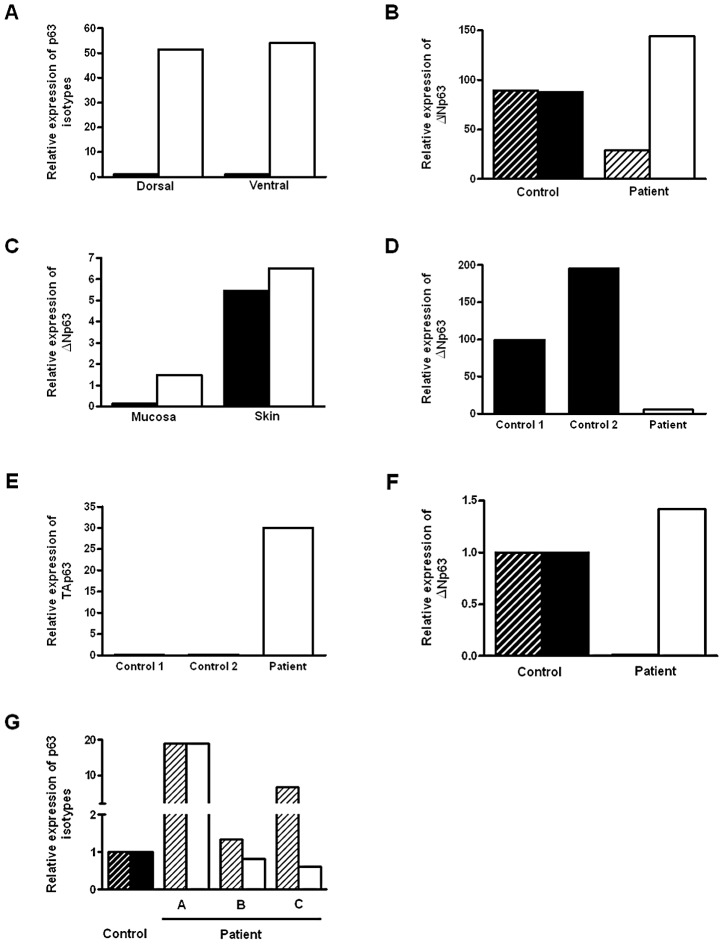
Figure 1. Expression of p63 isoforms in BEEC patient tissue and controls.
(A) Real-time qPCR of ΔNp63 (white column) and TAp63 (black column) expression dorsal and ventral foreskin from normal control (2 yr. male). (B) Real time qPCR of ΔNp63 expression dorsal (black hatched column) and ventral foreskin (black column) from 2 normal controls and dorsal (hatched column) and ventral foreskin (white column) from a BEEC patient. (C) Real-time qPCR of ΔNp63 expression in from BEEC patient (8 yr. male) dorsal (black column) and ventral (white column) foreskin mucosa and skin. (D) Real-time qPCR showing reduced ΔNp63 mRNA expression in BEEC bladder epithelium compared with 2 normal controls. (E) Increased TAp63 mRNA expression in BEEC bladder epithelium compared with 2 normal controls. (F) Real-time qPCR showing reduced ΔNp63 mRNA and increased TAp63 mRNA expression in bladder mucosa from a Caucasian BEEC patient (<1 yr. male) compared with normal controls (normalized to 1). (G) Real-time qPCR showing ΔNp63 and TAp63 mRNA expression in bladder mucosa from a three Caucasian BEEC patients (A, 13 yr. male; B, 14 yr. female; C, 6 yr. male) compared with normal controls (normalized to 1).

