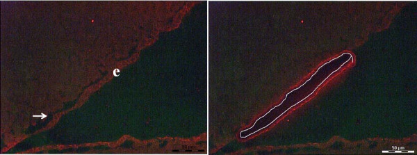
Figure 1.

Immunofluorescence labeling of ependymal cells. (A) Immunofluorescence labeling of cryostat sections from snap frozen brain tissue for ependymal cells (e) using anti- β catenin antibody followed by serial dehydration (B). Same section after subjected to LCM-mediated ependymal cells isolation.
