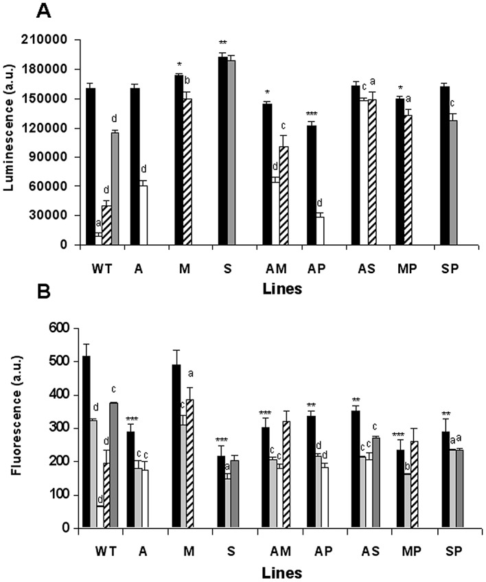
Figure 4. Effect of anti-leishmanial drugs on ATP synthesis and ΔΨm in resistant L. donovani promastigote lines.
(A) ATP levels were measured using CellTiter-Glo. (B) ΔΨm was measured by determining the accumulation of Rh123 (0.5 µM) in WT and resistant lines. In both cases, promastigote log-phase cultures were left untreated (controls, black columns) or exposed to 0.2 µM AmB (white columns), 25 µM MLF (oblique lines columns) for 3 h, 2 mM SbIII (gray columns) for 8 h, or 10 µM FCCP as a depolarization control (light gray columns) for 10 min. Measurements are expressed in arbitrary luminescence (panel A) or fluorescence (panel B) units ± SD from three independent experiments and show significant differences (a: p<0.05; b: p<0.01; c: p<0.005; d: p<0.001) when comparing the corresponding control values for each line with itself after treatment. A comparison of the different untreated lines with untreated WT lines also shows significant differences (*: p<0.05; **: p<0.005; ***: p<0.001).

