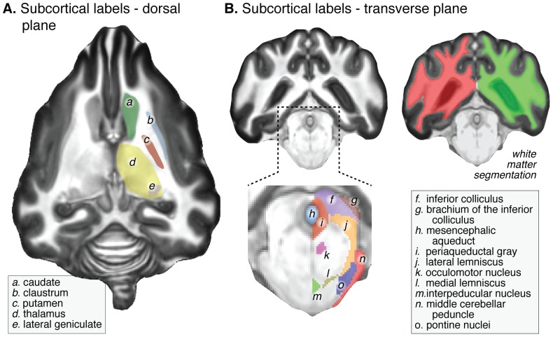
Figure 2. Representative slices of the high resolution ex-vivo template demonstrating labeled cortical and subcortical structures.
(A) Dorsal plane (horizontal) slice through the basal ganglia and thalamus. The fine structure of both the lateral geniculate nucleus and head of the hippocampus can be seen in this population average image. (B) Expanded and contrast-enhanced coronal slice through the brainstem, and illustration of white matter tissue segmentation.

