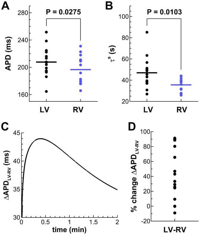
Figure 3. Functional LV/RV differences in the in vivo human heart.
A,B: Unipolar electrograms revealed longer steady-state APDs at the study CL and slower APD adaptation dynamics in LV compared to RV. C: Transient increased LV/RV APD dispersion following rate acceleration in a representative patient (Patient 3). D: Percent of ΔAPDLV-RV increase following rate acceleration for all patients of the study.

