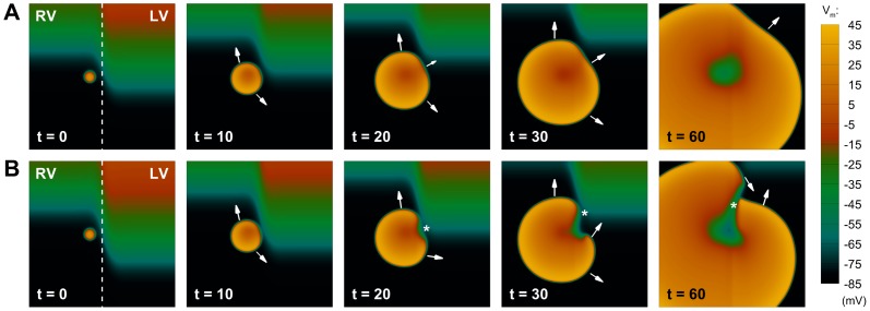
Figure 6. Development of unidirectional block due to transient patterns of interventricular APD dispersion.
A: Under average conditions of slow APD adaptation (scenario A), the transient APD dispersion between both ventricles only affects wavefront propagation partially, and the ectopic stimulation excites the whole tissue as a regular beat. B: For conditions of protracted slow APD adaptation (scenario B), a larger interventricular APD dispersion is able to produce unidirectional block (t = 20, marked by an asterisk), leading to the initiation of reentry (t = 30), that subsequently develops in the tissue (t = 60). Colorbar denotes transmembrane potential (mV); times indicated since initiation of ectopic stimulation (ms).

