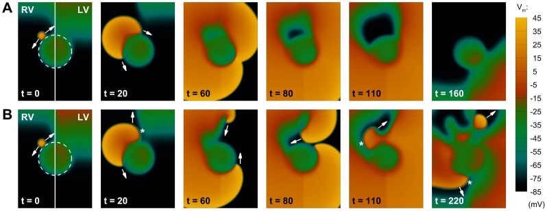
Figure 7. Interaction of transient patterns of interventricular APD dispersion with structural defects of the tissue.
The dashed line indicates an inexcitable region in the LV/RV junction. A: Under average conditions of slow APD adaptation (scenario A), interventricular APD dispersion is not able to produce conduction block, and the extra-stimulus proceeds circumventing the inexcitable area. B: For conditions of protracted slow APD adaptation (scenario B), the top part of the extra activation now finds a region of unidirectional block due to a larger APD dispersion (t = 20, marked by an asterisk). The wavefront therefore moves upwards, eventually developing into a reentrant wave (t = 60). Since the bottom part of the excitation has been circumventing the obstacle, the top reentrant wave can now proceed in the tissue (t = 80), finding new areas of conduction block (t = 110), and finally producing wave-break (t = 220). Figure annotation as in Figure 6.

