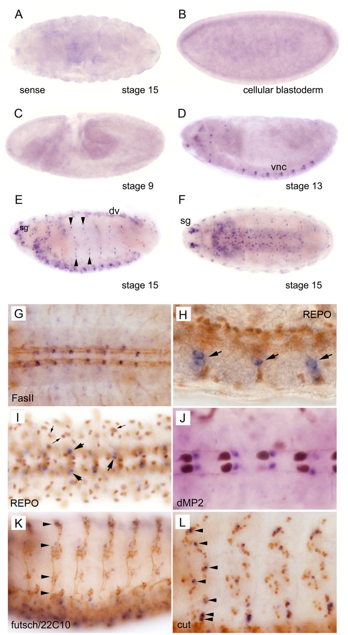
Figure 5. In situ hybridization analysis of DmGfrl expression during embryogenesis.
Cellular blastoderm (B) and early stage embryos up to stage 10 (C) do not express DmGfrl. Expression (blue staining) first appeared in the seven abdominal segments in the ventral nerve cord (D, vnc) and in unidentified ganglia in the head region at about stage 13. The expression became more widespread in the central and peripheral nervous system through the later stages of embryogenesis (E, F). DmGfrl was expressed in single cells or clusters of a few cells in the head sensory ganglia (E, sg), ventral nerve chord (D–F) and lateral sensory ganglia (E, arrowheads). At stage 15, dorsal vessel (dv) also expressed DmGfrl (E). Control hybridization with a sense probe did not show specific staining (A). (G–L) Following whole mount in situ hybridization, the embryos were subjected to immunoperoxidase staining (brown color) with neuronal and glial marker antibodies. Co-staining for the neuronal marker FasII showed that DmGfrl-expressing cells are localized within the VNC, along the longitudinal axon bundles (G). A lateral view on the VNC shows that the DmGfrl signal did not colocalize with the nuclear staining for REPO, a glial cell marker (H, arrowheads show DmGfrl+ cells). A ventral view of the VNC also shows the paired DmGfrl+ cells in each segment do not co-localize with REPO (I, large arrows show DmGfrl+ cells). However, in the more lateral and dorsal cells there may be some overlap in the signals for DmGfrl and REPO (I, small arrows). The DmGfrl+ cells localized posteriorly from the dMP2 interneurons (J, brown staining) in late-stage embryos, indicating that they are not dMP2 neurons and likely not vMP2 neurons either. Futsch/22C10 staining visualizes that the lateral axonal projections along which the DmGfrl+ cells were located (K, arrows). Staining for cut, a sensory neuron marker showed that the DmGfrl-expressing cells were within the external sensory organ cell clusters and likely all positive for cut (L, arrows). Original magnification was 400X in h, i and l, and 200X in all other images.

