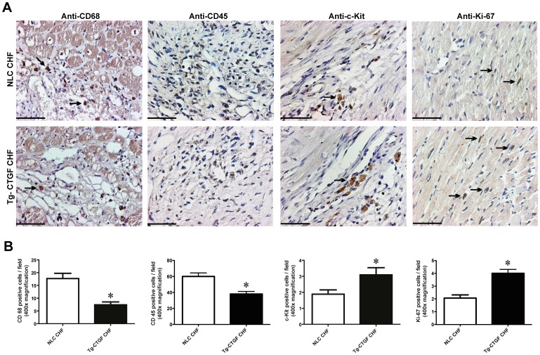
Figure 7. Morphometric analyses of inflammatory cells and cells undergoing apoptosis in myocardial tissue after MI.
Photomicrographs of immunohistochemical staining of CD68, CD45, c-Kit and Ki-67 in myocardial sections of hearts from NLC mice and Tg-CTGF mice 4 weeks after MI. Panels are from border zone of MI. Size bar indicates 50 µm. Arrows indicate examples of immunoreactive cells. Magnification: ×400. B. Histograms of CD68+-cells, CD45+-cells, c-kit+-cells and Ki-67+-cells (immunoreactive cells/400x power field) in peri-infarct region of NLC CHF and Tg-CTGF CHF mice 4 weeks after ligation of LAD. 5 visual fields/section and 3 sections per mice were analyzed. Data are mean±SEM of Tg-CTGF CHF (n = 4) and NLC CHF mice (n = 4). *P<0.05 vs. NLC CHF group.

