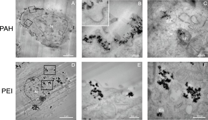
Figure 3. TEM observations of the distribution in NIH/3T3 EWS-Fli1 cells of ND-PAH (A)–(C) and ND-PEI (D)–(F) after 4 h of incubation.
N: cell nucleus, En: endosomal compartment, Ly: lysosomal compartment, Mp: macropinosome compartment. (A) Most of ND-PAH are located on the outside of the cell membrane. (B) Higher magnification of the cell membrane (upper square-delimited region of (A)) displaying invaginations around aggregates of ND-PAH, an early stage in clathrin-mediated endocytosis. Inset: Higher magnification showing the clathrin pits, perpendicular to the invaginated membrane. (C) Magnification corresponding to the lower square-delimited region in (A), showing ND-PAH located in endosomal (En) and lysosomal (Ly) vesicles. (D) Most of the ND-PEI are present within the cell, in large structures in the perinuclear region. (E) Magnification corresponding to the upper square-delimited region in (D) showing pseudopods formed at the cell membrane during ND-PEI uptake. (F) Magnification corresponding to the lower square-delimited region in (D), showing ND-PEI localization, mostly in large macropinosome (Mp) vesicles, but also, to some extent, in endosomes (En). Scale bars in (A) and (D): 2 µm and in (B), (C), (E) and (F): 200 nm. TEM magnification in (A) and (D): ×4,400; in (B) and (C): ×50,000; in (E): ×20,000, and in (F): ×30,000.

