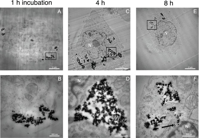
Figure 4. TEM imaging of the distribution of ND-PEI in NIH/3T3 EWS-Fli1 cells as a function of time N: cell nucleus, Mp: macropinosome compartment.
The cell were incubated with ND-PEI for 1 h (A)&(B), 4 h (C)&(D) and 8 h (E)&(F). (A) ND-PEI were rapidly taken up by cells: at 1 h, some NDs had already crossed the cell membrane. (B), (D) and (F) are magnifications corresponding to the regions surrounded by rectangle or square in (A), (C) and (E) respectively. (B) Pseudopod formation at the cell membrane during ND-PEI uptake. At 4 h (C) and 8 h (E), all the ND-PEI are located in large perinuclear vacuoles within the cell. Most of these structures correspond to macropinosomes, which are still delimited by a membrane at 4 h (D), but are disrupted after 8 h of treatment, as confirmed by the presence of free diamond nanocrystals in the cytoplasm (arrow)(F). Scale bars in (A), (C) and (E): 2 µm and in (B), (D) and (F): 500 nm. TEM magnification in (A), (C) and (E): ×4,400; in (B): ×20,000; in (D) and (F): ×30,000.

