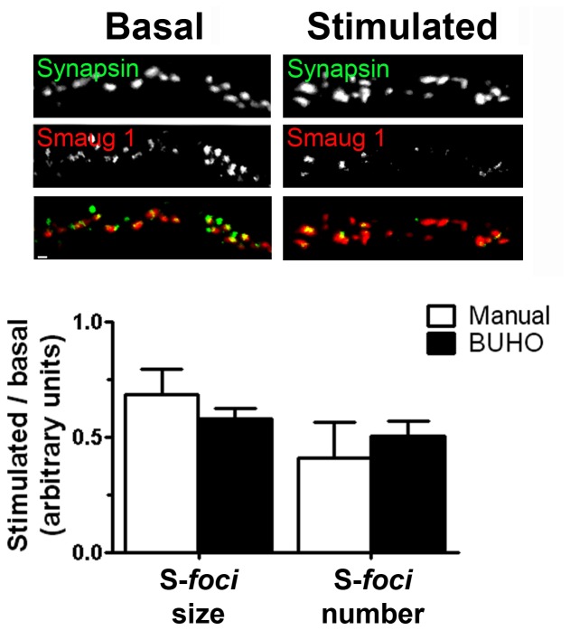
Figure 4. S-foci dissolution upon synaptic stimulation.
Cultured rat neurons were stimulated with NMDA and S-foci and synapses were identified with specific antibodies as indicated in Materials and Methods. The S-foci size and the number of synapses containing S-foci in their surroundings at a distance lower than 0.5 µm were evaluated. S-foci smaller than 0.2 µm2 were not included. Six micrographs (63×, 1024×1024 pixels), containing 4180 synapses were analyzed using BUHO, and a subset of 200 representative synapses were analyzed manually. Values relative to basal conditions are plotted. Error bars indicate standard deviation. Size bar, 2 µm.

