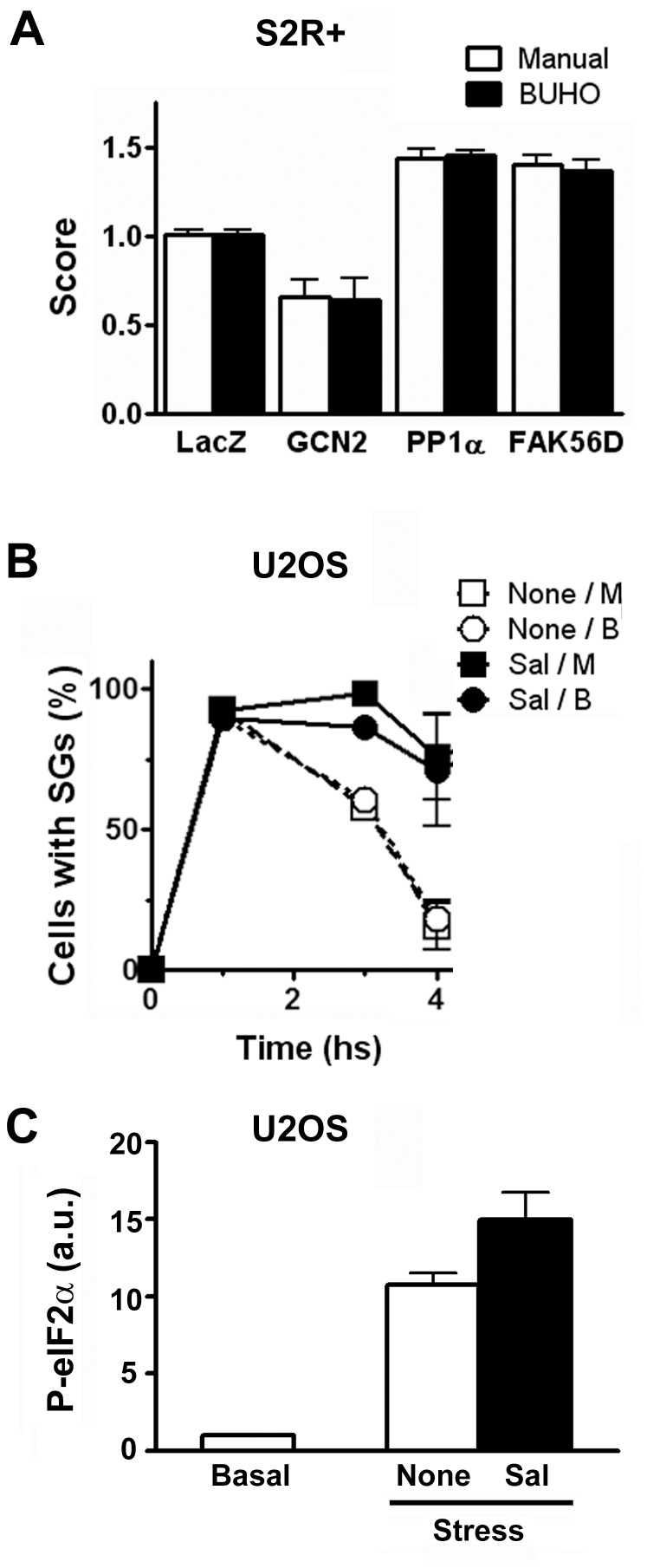
Figure 5. PP1α governs SG disassembly.
A, Drosophila S2R+ cells were exposed to the indicated dsRNA and the effect on SG formation was evaluated. Triplicate wells of the indicated RNAi treatments were analyzed both manually and by BUHO. 204 control wells were analyzed by BUHO, and a subset of 6 wells (see Table 3) was analyzed manually. The score is the ratio of the percentage cells with SGs in each well relative to the average percentage of cells with SGs in the control wells in the same plate. GCN2 knockdown impaired SG formation, and the KD of FAK56D or PP1α-96A facilitated their assembly. Scores determined manually differ in less than 7% from those calculated by BUHO. B and C, Mammalian U2OS cells were exposed to ER-stress in the presence (Sal) or absence (none) of salubrinal, as indicated in Materials and Methods. B, The number of SG-positive cells at the indicated time points was evaluated manually (M) or by BUHO (B) in 63×, 1024×1024 images. Error bars, standard deviation. C, Phosphorylation of eIF2α relative to basal conditions was evaluated three hours after stress induction in the presence or absence of salubrinal, as indicated in Materials and Methods. Error bars, standard deviation.

