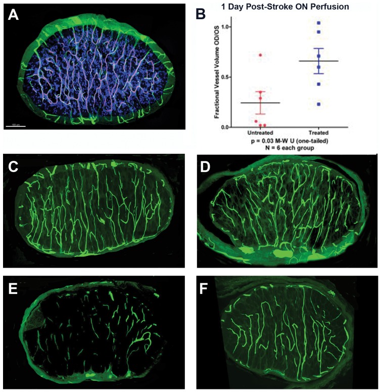
Figure 2. Microvascular analysis of naïve and infarcted ONs at 4 hours and 1 day post-induction.
A. FITC-BSA vascular imaging of naïve ON. Filled capillaries are relatively uniformly distributed throughout the nerve, communicating with peripheral vasculature. B. Quantitative capillary analysis of 1 day post-rAION-induced ON, in vehicle- and PGJ2-treated animals. Infarcted nerves were compared with the contralateral (naïve control) nerve of the same animal (1.0 of filling), and expressed on the Y axis as a fractional vessel volume (OD/OS). PGJ2-treated animals show significantly more patent capillaries at one day than vehicle treated animals (Mann-Whitney U test, p<0.03). C and D: ON capillary filling 4 hours post-induction. There is minimal loss of capillary patency in both vehicle- (panel C) and PGJ2-treated (panel D) nerves. E and F: ON capillary filling 1 day post-induction. There is significant loss of vascular patency in ONs of vehicle treated animals (panel E). ONs in PGJ2-treated animals (panel F) reveal considerably more patent vasculature at one day. Scale bar: 100 microns.

