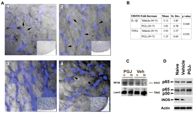
Figure 7. PGJ2 inhibits WM related NFκB activity.
A. Ni-DAB quenched fluorescence analysis of NFκB expression and nuclear localization. ON sections developed and incubated identically to analyze relative Ni-DAB staining. Panels 1 and 2: ON cross-sections without PGJ2 treatment. Panels 3 and 4: ON cross-sections following PGJ2 treatment. Panel insets: low power micrographs showing relative ON NFκB staining. Panel 1: NFκB expression in un-induced, vehicle-treated ON. Note reduced DAPI intensity (inset). NFκB is present in nuclei, which have reduced visibility (arrows). Panel 2: NFκB expression in vehicle treated ON 1 day post-rAION induction. Increased intracellular NFκB expression (inset), relative to naïve ON, with reduced DAPI-stained nuclear visibility (arrows). Panel 3: NFκB expression in uninduced ON 1 d post-PGJ2 treatment. Note increased Ni-DAB signal (compare inset with panels 1 and 2), prominent DAPI-stained nuclei and perinuclear NFκB staining. Panel 4: NFκB expression in rAION-induced ON 1 day post-PGJ2 treatment. Note increased NFκB signal, compared with vehicle- or naïve tissue (compare inset panel 4 with panels 1 and 2), increased perinuclear NFκB accumulation (arrowheads). B. NFκB-associated gene expression in infarcted ONs with and without PGJ2. Comparison of infarcted- and contralateral nerves, expressed as R/L ratios, treated with vehicle or PGJ2, individual ON results (n = 12). C. ON western blot analysis: NFκB-subunit expression 1 d post vehicle- and PGJ2-treated animals. p65 signal increased in uninfarcted (U) PGJ2-treated ON and PGJ2-infarcted (I) ON, compared with infarct controls. Loading control: Lamin C (LamC). D. Western analysis: NFκB subunit- and NFκB-related (iNOS) expression in CNS (corpus callosum). p65 subunit expression with p65-specific antibody (Santa Cruz). Increased p65 signal seen in PGJ2- compared with vehicle-treated animals. Second row: expression using antibody with overlapping p65/p50 subunit specificity. Increased p65 is independent of p50 subunit levels. Third row: iNOS expression. PGJ2 treatment results in decreased white matter iNOS protein expression, compared with naïve or vehicle-treated animals. Loading control: β-Actin.

