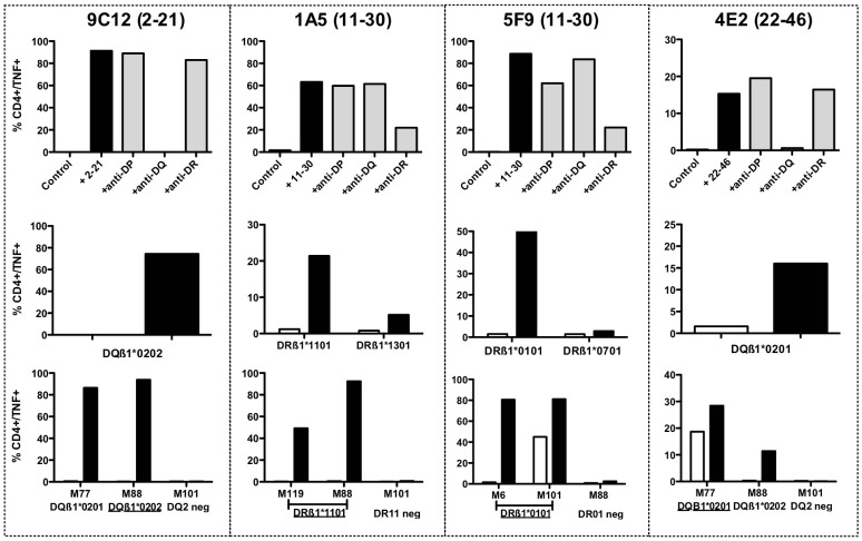
Figure 6. HLA-restricting element of MELOE-1 specific T cell clones and reactivity against HLA-matched melanoma cell lines.
Upper panel: the HLA restriction of MELOE-1 specific T cell clones was assessed by intracellular TNF labelling, using anti-HLA blocking antibodies. T cell clones were stimulated either with peptide alone (10 µM) in an autopresentation assay and in presence or not of blocking antibodies at a concentration of 12.5 µg/mL. Middle panel: HLA restriction was confirmed with HLA-matched B-EBV cell lines unloaded (white bars) or pulsed (black bars) 2 h with the cognate peptide, at an effector/ratio of 1/2. Lower panel: reactivity of each T cell clone against HLA-class II expressing melanoma cells (ratio 1/2) was assessed by intracellular TNF labelling, in presence (black bars) or not (white bars) of exogenous peptide.

