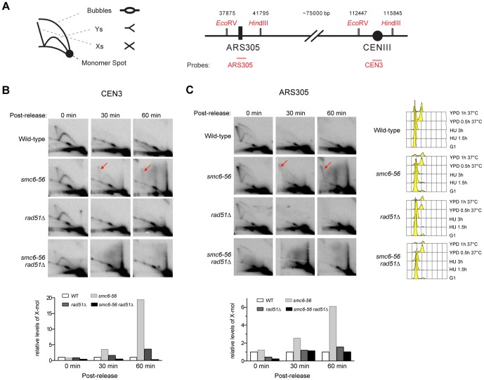
Figure 1. smc6-56 cells accumulate recombination intermediates at centromeric and ARS305 sequences.
(A) Schematics of 2D gel and genomic regions containing ARS305 and CEN3 sequences. The numbers above the genomic region are base pair coordinates from the left end of chromosome III. (B–C) Cells were arrested in G1 using alpha-factor, and synchronized in S phase using 0.2 M HU for 3 hours at 25°C. Cells were then washed and released into YPD medium at 37°C. Samples before and after release at indicated time points were examined by 2D gel analysis. Membranes were hybridized to a probe specific for the centromeric sequence on chromosome III (B) and another specific for ARS305 (C). FACS analysis before and after release is presented on the right panel in (C). Quantification of X-molecules (red arrows) is shown in the bottom panels. For both loci, the level of X-molecules increases in smc6-56 cells compared with wild-type, and rad51Δ suppresses these increases.

