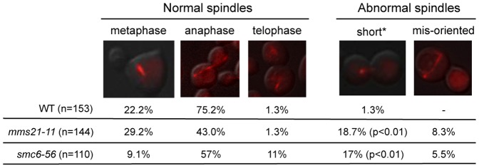
Figure 5. Spindle morphology in wild-type, mms21-11, and smc6-56 cells.
Spindle morphology was examined 75 minutes after cells were released from G1 arrest when the majority of cells were at anaphase. Only medium to large budded cells were counted. A representative picture is shown for each spindle category. Similar results were obtained for two strains of each genotype and the results of one pair are shown. Asterisk denotes large budded cells with short spindles. p value indicates that there is a statistically significant difference between wild-type and mutants.

