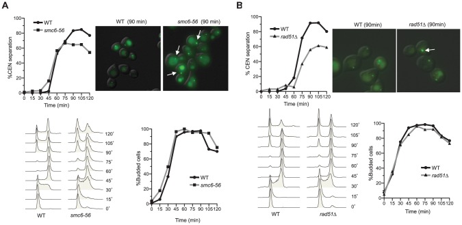
Figure 6. smc6-56 and rad51Δ cells are defective in centromeric LacO array separation.
(A–B) smc6-56 and rad51Δ cells exhibit defects in centromere separation. Cells contain a LacO array integrated 12 kb distal to the centromere on chromosome IV. Cells were arrested in G1 at 23°C and then shifted to 37°C for 1 hour before release into the cell cycle at 37°C. Samples were taken every 15 minutes to examine cell cycle progression by FACS analysis (bottom left) and budding index (bottom right). These time points were also examined for centromere separation by microscopy (top left). The difference between the percentage of wild-type and rad51Δ or between that of wild-type and smc6-56 cells containing separated GFP foci at 90, 105, and 120 min after release is statistically significant (p<0.01). Representative pictures at 90 minutes are shown (top right); arrows indicate cells with unseparated centromeres. Note that the background signals of LacI-GFP represent vacuolar staining.

