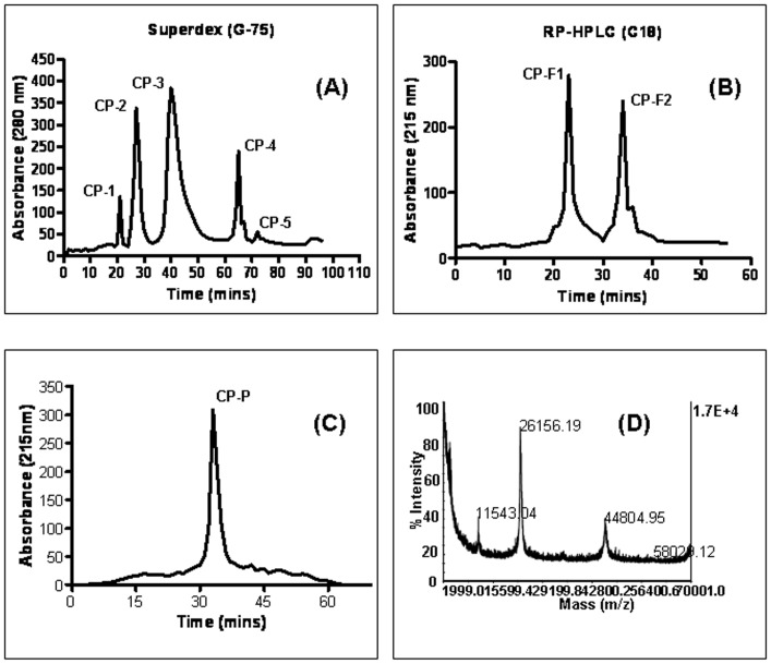
Figure 1. Purification and characterization of anticancer protein CP-P.
(A) The clear supernatant of Calotropis procera root-bark was separated by gel-filtration (Superdex G-75) chromatography into five major peaks CP-1–CP-5, (B) the most active peak (CP-3) was further separated by reverse-phase high performance liquid chromatography (RP-HPLC) by using a C18 column which produced two peaks namely CP-F1 and CP-F2, (C) the active fraction (CP-F1) was resolved by a C8 column and gave a single peak named Calotropis procera protein “CP-P”, (D) CP-P mass was analyzed by a perspective biosystem matrix-assisted laser desorption ionization-time of flight (MALDI-TOF/MS).

