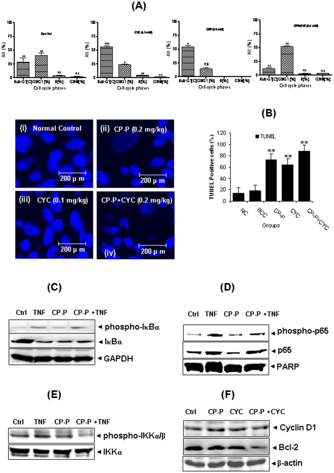
Figure 3. The number of apoptotic cells in breast tumor sections was detected by TUNEL assay.
(A) The apoptotic effect of CP-P alone or in a combination of CYC treatment was analyzed by flow cytometry to compare the various cell cycle phases. The CP-P alone, or in combination with CYC, induced promising apoptotic effects. As a result, fewer MCF-7 cells accumulated in the Sub-G1 phase compared to the control. (B) TUNEL positive cells were quantified, revealing that apoptotic cells in breast tumors increased significantly in CP-P or CYC drug alone, or in combination, groups versus untreated cells. CP-P and CYC drug-treated groups were compared to breast cancer control groups. The combination treatment induced more positive cells than others. Values are mean ± S.D. (n = 3) replicates, **P<0.01 values indicate significance to control. (C) CP-P inhibits TNF-α -induced nuclear degradation/phosphorylation of IκBα. MCF-7 cells were either untreated or pretreated with 50 µg/ml CP-P for 6 h at 37°C and then treated with 1 nM TNF-α for 30 min. Cytoplasmic extracts were prepared and analyzed by Western blotting using antibodies against anti- IκBα and phospho-specific IκBα. The same membrane was reblotted with GAPDH antibody to verify equal loading. The results shown are representative of two independent experiments. (D) CP-P inhibits TNF-α-induced nuclear phosphorylation/translocation of p65. MCF-7 cells were either untreated or pretreated with 50 µg/ml CP-P for 6 h at 37°C and then incubated with 1 nM TNF-α for 30 min. Nuclear extracts were prepared and analyzed by Western blotting using antibodies against anti-p65 and phospho-specific p65. The same membrane was reblotted with PARP antibody to verify equal loading. The results shown are representative of two independent experiments. (E) Effect of CP-P on TNF-induced phosphorylation of IKK-α and IKK-β. MCF-7 cells were incubated with 50 µg/ml CP-P for 6 h at 37°C and then treated with 1 nM TNF-α for 15 min. Whole cell extracts were prepared and analyzed by Western blot analysis using anti-phospho-specific IKK-α/β antibody. The same membrane was reblotted with anti-IKK-α antibody to verify equal loading. (F) CP-P inhibits TNF-α -induced expression of NF-κB-dependent proliferative and antiapoptotic proteins. MCF-7 cells were incubated with 50 µg/ml CP-P for 6 h at 37°C and then treated with 1 nM TNF-α for 24 h. Whole-cell extracts were prepared and analyzed by Western blot using the indicated antibodies (Cyclin D1 and Bcl-2). The same membrane was reblotted with β-actin antibody to verify equal loading. Results are representative of two independent experiments.

