
Figure 3. Representative pictures of MHC class I, ICAM-1 and Foxp3 expression using immunohistochemistry on tumor specimens of melanoma invaded LN.
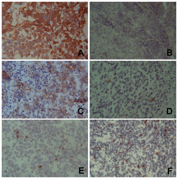
(A) and (B): MHC class I expression, (C) and (D): ICAM-1 expression, (E) and (F): Foxp3 expression (magnification×25). (A), (C) and (E) are specimens from the group with only one invaded LN whereas (B), (D) and (F) are specimens from the group with more than one invaded LN.