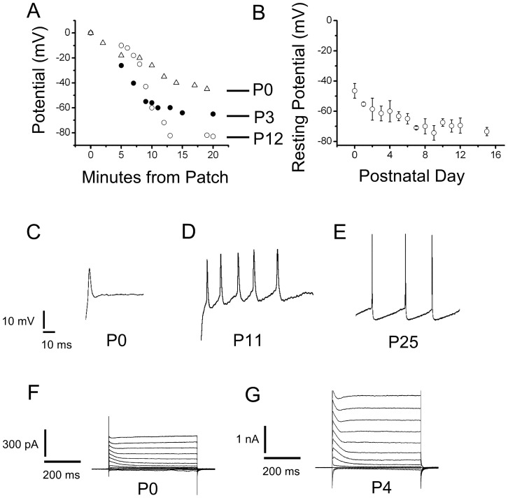
Figure 2. Excitability Changes in SN DA neurons during development.
(A) Resting potential measurements for three SN DA neurons as a function of time. After obtaining a cell-attached patch, we waited to gain access as the gramicidin diffused from the back of the pipette onto the tip, measuring the voltage drop necessary to maintain a zero current, in the search mode. The resting potential equilibrated slowly as gramicidin was incorporated into the patch. Only recordings that developed over the course of several minutes and were stable after reaching equilibrium were used. Open triangles, neuron at P0. Closed circle, neuron at P3. Open circles, neuron at P12. (B) Resting potentials measured with the method illustrated in A, are plotted as a function of postnatal day of development. (C),(D) & (E) Electrical activity of SN DA neurons in response to depolarizing current pulses. Neurons in the fast-current clamp mode, were depolarized by current injections. P0 to P5 neurons never displayed spontaneous firing. The P0 neuron shown here was depolarized to −20 mV, from a resting potential of −40 mV. The P11 neuron was depolarized to −40 mV from a resting voltage of −60 mV. The P25 could fire a continuous train of action potentials from resting potential of −60 mV. Small current injections were performed to adjust the resting voltage. Small current injections were performed from resting voltages (Notice difference between P0 and P11). This was on purpose to avoid large current injections that could distort the recording. However, even when depolarized from −60 mV, P0 and P1 neurons never fired action potential continuously, but rather the small wide action potentials shown here. (F) Typical voltage clamp current in responses to voltage pulses with 20 mV increments from a holding potential of −80 mV to test pulses from −120 to 100 mV with 20 mV increments. Raw traces shown, no leak subtraction. P0 neuron shows the typical outward current with an inactivating IA current and a non-inactivating current. Small I h and inward rectifier currents were also observed at hyperpolarized potentials less than −80 mV. (G) A P4 neuron with a larger outward current, in response to the same voltage protocol as in F. This neuron also had larger I h and inward rectifier currents, not analyzed here.

