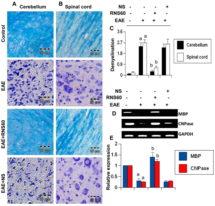
Figure 10. RNS60 inhibits demyelination in the CNS of EAE mice.
Cerebellar (A) and spinal cord (B) sections of control EAE (16 dpt), and either RNS60− or NS-treated EAE mice (16 dpt receiving RNS60/NS from 8 dpt) were stained with Luxol fast blue. Digital images were collected under bright field setting using a 40× objective. (C) Demyelination was represented quantitatively by using a scale as described in materials and methods. Data are expressed as the mean ± SEM of five different mice. ap<0.001 vs control; bp<0.001 vs EAE. Cerebellum of control, EAE and either RNS60− or NS-treated EAE mice was analyzed for MBP and CNPase by semi-quantitative RT-PCR (D) and quantitative real-time PCR (E). Data are expressed as the mean ± SEM of five different mice. ap<0.001 vs control; bp<0.001 vs EAE.

