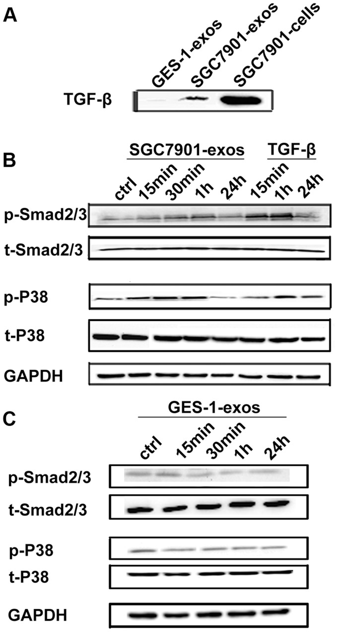
Figure 4. Gastric cancer cell derived exosomes induce Smad2/3 and p38 phosphorylation in hucMSCs.
A. Western blotting analyses of TGF-β expression in gastric cancer cell (SGC7901) and normal gastric epithelial cell (GES-1) derived exosomes. B. HucMSCs were treated with SGC7901 derived exosomes (800 µg/mL) for different times as indicated. The levels of p-Smad2/3, t-Smad2/3, p-p38and t-p38 were analyzed by Western blotting. TGF-β served as a positive control. C. HucMSCs were treated with GES-1 derived exosomes (800 µg/mL) for different times as indicated. The levels of p-Smad2/3, t-Smad2/3, p-p38 and t-p38 were analyzed by Western blotting.

