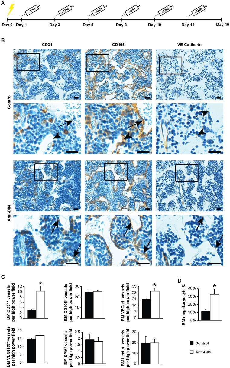
Figure 1. Therapeutic anti-Dll4 blockade interferes with the BM vascular niche.
(A) Schematic representation of the clinical assessment of anti-Dll4 treatment. Yellow lightening bolt, sub-lethal irradiation. (B) Immunohistochemistry for CD31, CD105 and VE-Cadherin counterstained with Mayer’s haemalum (Leica DMD 108). Sequential sections represent the same blood vessels. Arrowhead, CD31-CD105+Ve-Cadherin+ blood vessel; dashed arrow, CD31−CD105+VE-Cadherin− blood vessel; arrow, CD31+CD105+VE-Cadherin+ blood vessel. Bar = 20 µm. (C) CD31, CD105, VE-Cadherin, VEGFR3, SMA and Lectin-positive vessel count, per high power field (400x, Leica DMD 108), reveal an increase of CD31 and VE-Cadherin-positive BM vessels in anti-Dll4 treated mice. (D) Flow cytometric analysis of the percentage of megakaryocytes (CD41+ cells) in the BM shows an increase of BM megakaryocyte cell percentage in anti-Dll4 treated mice. Data are means ± s.e.m. *, p<0.05; data represents one of three experiments in which n = 3.

