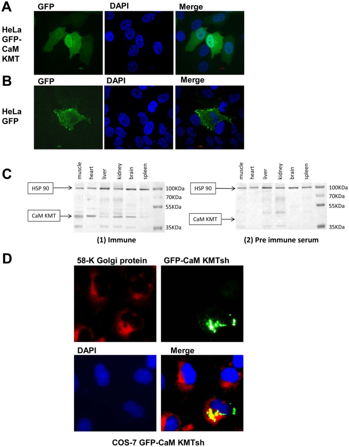
Figure 3. Subcellular localization of the CaM KMT-GFP fusion proteins in transiently transfected cells and expression in mouse tissues.
(A) GFP- CaM KMT is localized in the cytoplasm and the nucleus. Confocal images of HeLa cells expressing CaM KMT-GFP (green), nuclear staining by DAPI (blue) and the merged image. (B) The expression of the GFP only. Confocal images of HeLa cells expressing GFP (green), staining of nuclei by DAPI (blue), and the merged image. (C) Cell lysates (100 µg of protein/lane) from mouse muscle, heart, liver, kidney, brain and spleen were resolved by SDS-PAGE, transferred to a nitrocellulose membrane, and blotted with an affinity purified polyclonal anti-CaM KMT antibody (1) immune and (2) pre-immune serum. Anti-HSP90 antibody served for protein loading control, 100 µg protein/lane were analyzed. Positions of CaM KMT and HSP-90 are indicated by the arrows. (D) GFP- CaM KMTsh is localized to the Golgi. COS-7 cells were transfected with the GFP- CaM KMT short variant and immunostained with primary antibodies against Golgi 58 k protein. GFP-CaM KMTsh was detected directly by the fluorescence microscopy (green) and 58 k Golgi protein was visualised with Cy3-labeled secondary antibodies (red). Cells nuclei were stained with DAPI (blue). Shown is the merged image presenting colocalization (in yellow) of the GFP-CaM KMT short protein with Golgi apparatus.

