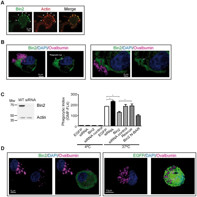
Figure 6. Bin2 is located at the phagocytic cup of macrophages and regulates phagocytosis. A .
: Epifluorescence micrographs of fixed macrophages (mouse BAC1·2F5 cell line) transiently overexpressiong Bin2-EGFP and LifeAct-mCherry. Cells were incubated with 0.9 µm latex beads for 3 min. before fixation to be able to visualize phagocytic cups (white stars). B: Confocal micrographs of two different rat alveolar macrophages (RAM) over-expressing Bin2-EGFP. Cells were incubated with alexa-labelled immune-complexed ovalbumin (pink) for 10 minutes at 37°C. Nuclei stained with DAPI (blue). Bin2 is enriched at the phagocytic cup. C: Left: western blot of RAM lysates from wild type (WT) and 48h-rBin2 siRNA transfected cells developed with polyclonal anti-hBin2 (BACT) and anti-actin (loading control) antibodies. Molecular weight markers (Broad Range, Promega) are indicated. Right: phagocytosis assay (expressed as phagocytic index (geometric mean fluorescence of positive cells)) performed with RAM cells at 4° (surface binding without internalization) and 37°C (internalization). Cells were incubated with alexa-labelled ovalbumin for 45 min before analysis by flow cytometry. Uptake from cells overexpressing rBin2-EGFP (Bin2) and rBin2 N-BAR-EGFP (N-BAR) and Bin2-depleted cells (Bin2 siRNA) where compared with control experiment (EGFP). Overexpression of a siRNA-insensitive protein on Bin2 depleted cells rescues the phenotype (Rescue). A siRNA control was also performed (siRNA control). Data are the mean ± SD. Significance was calculated using the Student’s t test (* = p<0.05). D: Confocal micrographs of RAM cells over-expressing Bin2-EGFP (Bin2, green, left) or control (EGFP, green, right). Cells were incubated with alexa-labelled immune-complexed ovalbumin (pink) for 120 minutes 37°C. Nuclei stained with DAPI (blue).

