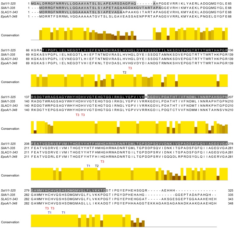
Figure 1. Multiple sequence alignment of homologue small laccases.
Ssl1 from Streptomyces sviceus, SilA from S. ipomoea, EpoA from S. griseus and SLAC from S. coelicolor. The copper binding residues are conserved in all 4 laccases as indicated. All four laccases consist of 2 domains (indicated in Ssl1, black: domain 1, dark grey: domain 2). Signal sequences of the twin-arginine pathway are indicated in light grey. Sequence identity between the four laccases is high and variations are mainly located at the termini.

