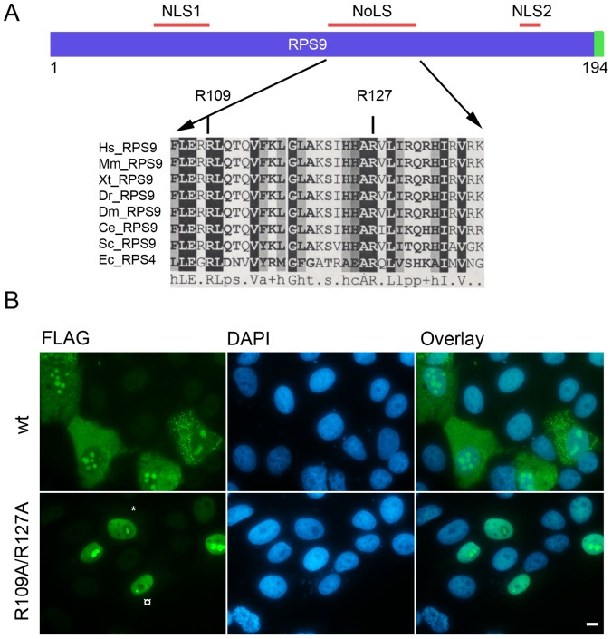
Figure 5. Altered localization of RPS9-FLAG NoLS domain mutant.
A.) Schematic representation of RPS9 with the NLS1, NLS2 and NoLS. Below is a partial multiple sequence alignment of RPS9/RPS4 depicting the central motif that confers nucleolar localization and with the conserved arginine residues mutated to alanine (R109A) and (R127A) shown. Abbreviations: Hs- Homo Sapiens, Mm – Mus Musculus, Xt- Xenopus Tropicalis, Dr – Danio Rerio, Dm – Drosophila Melanogaster, Ce- Caenorhabditis Elegans, Sc – Saccaromyces Cerevisiae, Ec- Escherichia Coli. B.) The combined mutation of R109 and R127 residues to alanine disrupted the cytoplasmic/nucleolar staining pattern seen in wt RPS9-FLAG expressing cells. A subset transfected cells displayed a more intensely stained sub-nucleolar structure as is indicated with an asterix (*) in the figure, while other transfected cells had such structures in the nucleolar proximity as indicated with the (¤) symbol. Plasmids were transfected transiently into U2OS cells and stained for FLAG expression 24 hours after transfection. Bar 10 µM.

