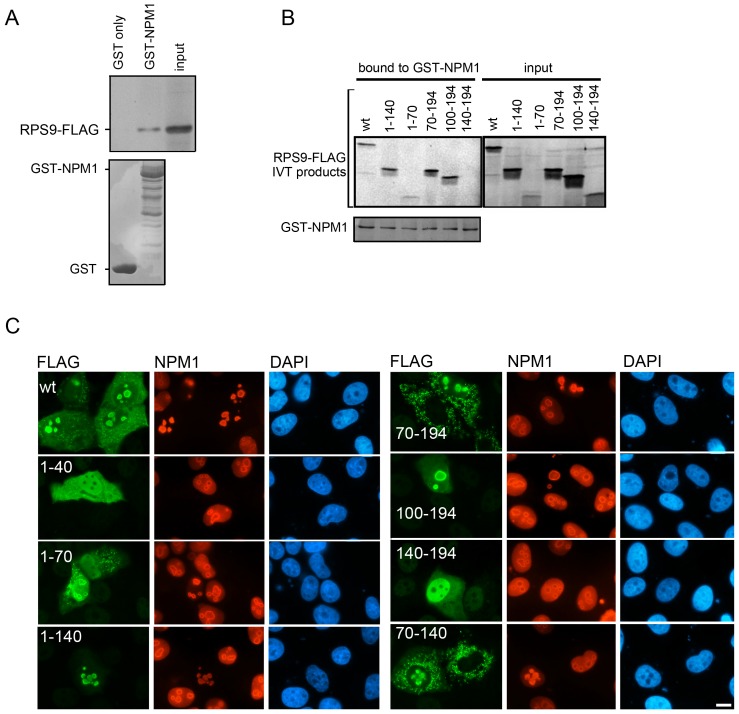
Figure 6. Binding and co-localization of different RPS9 domains with NPM1.
A.) In vitro translated RPS9-FLAG binds to GST-NPM1 but not GST. RPS9-FLAG was incubated with GST or GST-NPM1 on beads and bound product was visualized by autoradiography (30% of input is shown). B.) In vitro translated RPS9-FLAG deletion mutants and purified GST-NPM1 revealed that fragments 1–70, 1–140, 70–194, and 100–194 but not RPS9140–194-FLAG were able to bind GST-NPM1. The binding of radiolabeled RPS9-FLAG to GST or GST-NPM1 was detected by autoradiography (20% of input is shown). Coomassie brilliant blue staining of the gel demonstrated equivalent levels of input GST. C.) Co-localization of NPM1 and RPS9-FLAG. U2OS cells were transfected with wt RPS9-FLAG and RPS9-FLAG deletion mutants as indicated in the figure. Cells were double stained for FLAG and NPM1 expression using a rabbit polyclonal anti-FLAG antibody and an NPM1 monoclonal antibody. Bar 10 µM.

