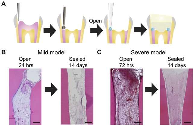
Figure 1. The establishment of mild and severe irreversible pulpitis models.
A. Schematic diagrams of the amputation of the dog mature molar pulp tissues and subsequent cavity sealing. The crowns of the upper and lower premolars were removed, and the pulp tissues were amputated using a round burr. The amputated pulp tissues were exposed to allow for infection and treated with solution absorbed in Spongel. After the treatments, the cavity were sealed with phosphate cement and light-cured composite resin. B. The histology of the pulp tissues from dogs with mild irreversible pulpitis. The left panel shows amputated pulp tissue that remained exposed for 24 hours. After 24 hours, the cavity was covered with spongel and sealed. The right panel shows the pulp at 14 days after sealing. C. The histology of the pulp tissues from dogs with severe irreversible pulpitis. The left panel shows amputated pulp tissue that remained exposed for 72 hours. After 72 hours, the cavity was sealed. The right panel shows the pulp tissue at 14 days after sealing. Scale bar, 500 µm.

